To alleviate
the pain of a ruptured or herniated intervetebral disc, a Laminotomy/Microdiscectomy may be performed.
This surgical procedure
is carried out in two steps beginning with the laminotomy. Lamina is the Latin name given to the bone protecting the spinal canal, and otomy means opening or hole. The laminotomy simply opens up the spinal canal
in order to visualize the pinched nerve root.
Once this is accomplished,
the second procedure, the microdiscectomy, is performed. A high
powered stereoscopic microscope is used to provide illumination
and magnification to allow the nerve and surrounding structures to be
visualized clearly through an incision less than one inch long.
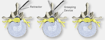
The nerve root is carefully protected with a specialized retractor, and
protruding disc fragments, along with any remaining loose or degenerated
disc material, are then removed with a small grasping device.
The small
hole left in the annulus will regenerate in 4 to 6 weeks and fill
in with new disc material.